
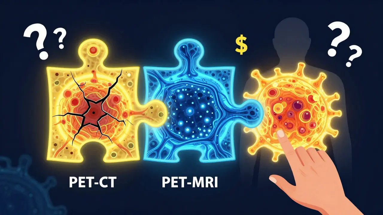
When it comes to fighting cancer, knowing exactly where the disease is-and how far it’s spread-isn’t just helpful, it’s life-or-death. That’s where oncologic imaging comes in. Three technologies dominate this space: PET-CT, MRI, and the newer hybrid PET-MRI. Each has its strengths, its limits, and its sweet spot in cancer care. There’s no single best scan for every patient. The right choice depends on the type of cancer, the clinical question, and even the hospital’s resources.
PET-CT: The Workhorse of Cancer Staging
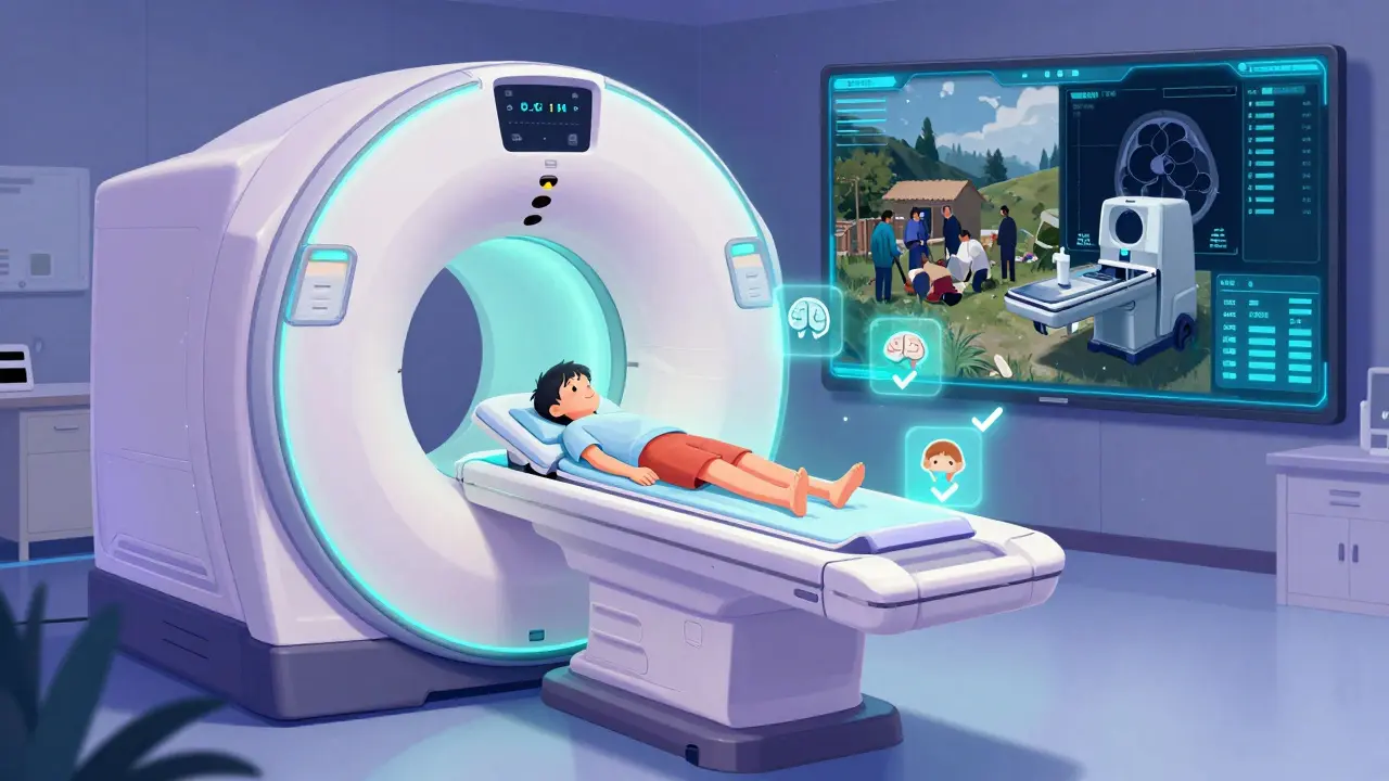
PET-CT became the standard for cancer staging after its FDA approval in 2001. It combines two scans into one: a PET scan that shows metabolic activity (using a radioactive sugar tracer called 18F-FDG) and a CT scan that maps anatomy in sharp detail. Think of it like this: the PET part tells you where the cancer is active, and the CT part tells you where it’s physically located.
It’s fast. A typical scan takes 15 to 20 minutes. It’s widely available. Most hospitals have one. And it’s proven. For cancers like lung, lymphoma, and colorectal cancer, PET-CT is often the first choice for staging. A 2023 meta-analysis found it correctly identified cancer spread in 84% of non-small cell lung cancer cases. It’s also excellent for spotting distant metastases-like tumors in the liver or bones-that might not show up on other scans.
But it has downsides. It uses ionizing radiation-about 10 to 25 mSv per scan, which is roughly 3 to 8 times a standard chest CT. That matters for younger patients or those needing repeated scans. It also struggles with cancers that don’t absorb much FDG, like some prostate or low-grade tumors. And because CT uses X-rays, it can miss subtle soft tissue changes, especially in the brain, liver, or pelvic organs.
MRI: The Detail Master Without Radiation
MRI doesn’t use radiation. Instead, it uses powerful magnets and radio waves to create incredibly detailed images of soft tissues. That’s why it’s the gold standard for brain tumors, spinal cord cancers, liver lesions, and pelvic cancers like prostate, cervical, and rectal cancer.
Modern 3T MRI systems can resolve structures as small as 0.5 mm. Functional techniques like diffusion-weighted imaging (DWI) and dynamic contrast-enhanced MRI can reveal how water moves through tissue or how blood flows into tumors-clues that help distinguish cancer from scar tissue or inflammation. For example, in breast cancer, MRI can detect small tumors that mammograms miss, especially in dense breast tissue.
A 2022 study in Cancer found conventional MRI detected prostate cancer with 75% accuracy, compared to 62% for PSMA PET-CT. That’s a big deal when deciding whether a biopsy is needed. MRI is also unmatched in evaluating treatment response. After radiation or chemo, it can tell if a lesion is dead tissue or still alive-something PET-CT often can’t do reliably.
The catch? Time. An MRI can take 30 to 60 minutes. It’s noisy. It’s claustrophobic for some. And patients with pacemakers, certain metal implants, or severe kidney disease can’t have it. Plus, MRI doesn’t show metabolic activity the way PET does. So while it’s brilliant at showing structure, it doesn’t always tell you if a suspicious area is actively cancerous.
PET-MRI: The Hybrid Advantage
PET-MRI, first commercially available in 2011, merges the metabolic power of PET with the soft-tissue clarity of MRI-all in one machine. It’s not just a combo-it’s a synergy. The PET tracer lights up active cancer cells, and the MRI shows exactly how they’re nestled in organs, nerves, or blood vessels.
This matters most where detail is critical. In brain tumors, PET-MRI can tell the difference between tumor recurrence and radiation damage with 85-90% accuracy-far better than either scan alone. In liver cancer, a 2022 survey of radiologists found 68% reported higher diagnostic confidence with PET-MRI than PET-CT. For pediatric cancers, where reducing radiation is a priority, PET-MRI cuts exposure by nearly half compared to PET-CT.
It’s also changing the game in pelvic cancers. A 2023 RadioGraphics study showed PET-MRI led to changed treatment plans in nearly half of pancreatic cancer patients-because it spotted hidden metastases that PET-CT missed.
But PET-MRI isn’t perfect. It’s slower-45 to 60 minutes. It’s expensive. A single scan costs $2,500 to $3,500 in the U.S., compared to $1,600-$2,300 for PET-CT. The machines themselves cost $3-4.2 million, versus $1.8-2.5 million for PET-CT. And the technical challenges are real. Getting accurate PET images inside a strong magnetic field is hard. Attenuation correction artifacts (where the scanner misreads tissue density) occur in 63% of sites, requiring specialized physics teams to fix.
Which Scan Is Right for Which Cancer?
There’s no one-size-fits-all. Here’s how experts use them:
- Lung cancer: PET-CT remains first-line for initial staging. PET-MRI is reserved for complex cases where brain or adrenal involvement is suspected.
- Prostate cancer: PSMA PET-CT is excellent for detecting spread beyond the prostate. But for local staging-checking if cancer is still inside the gland-multiparametric MRI is superior.
- Breast cancer: For early response to chemo, PET-CT has higher specificity than MRI. But for dense breasts or high-risk patients, MRI is better for initial detection.
- Brain tumors: PET-MRI is now preferred. It distinguishes recurrence from radiation necrosis with far greater accuracy than either scan alone.
- Pancreatic and liver cancers: PET-MRI changes management in nearly half of cases. It’s becoming standard in major centers.
- Pediatric and young adult cancers: PET-MRI is increasingly favored due to lower radiation exposure and better soft tissue contrast.
Real-World Challenges
Even the best technology fails if it’s not used right. Radiologists report that PET-MRI requires 40+ extra hours of training compared to PET-CT. Technologists say motion artifacts during long scans-especially in the abdomen-are a common problem. One radiologist on Reddit noted, “We had a 14-year-old with neuroblastoma who couldn’t stay still. The scan had to be repeated twice.”
Reimbursement is another hurdle. Many insurance plans still don’t cover PET-MRI unless PET-CT was inconclusive. A 2021 study found 45% of cancer centers struggled with billing, even though 82% said the diagnostic value justified its use.
And availability? Only 1 in 5 hospitals in the U.S. has a PET-MRI scanner. Most are in academic centers. In rural areas or smaller clinics, PET-CT is still the only option.
The Future: AI, New Tracers, and Personalized Imaging
The next leap isn’t just better machines-it’s smarter interpretation. At the 2023 RSNA meeting, 27 presentations focused on AI-driven radiomics: using algorithms to pull hidden patterns from PET and MRI data to predict how a tumor will respond to treatment before it even starts.
New tracers are expanding what we can see. PSMA for prostate cancer, Ga-68 DOTATATE for neuroendocrine tumors, and F-18 fluciclovine for recurrent prostate cancer are now FDA-approved and being integrated with MRI. The NCI’s PREDICT trial is testing whether AI can combine these images to create personalized treatment plans.
Siemens Healthineers’ new BioMatrix 600 PET-MRI, cleared in January 2024, cuts scan time to just 6 minutes for a whole-body scan. That’s a game-changer for patient comfort and throughput.
By 2035, experts predict PET-MRI will hold 25-30% of the market in academic centers-not because it replaces PET-CT, but because it’s the best tool for specific, high-stakes decisions.
Final Takeaway
There’s no “best” imaging tool in oncology. PET-CT is fast, widely available, and great for most cancers. MRI gives unmatched detail without radiation-ideal for brain, liver, and pelvic tumors. PET-MRI is the future for complex cases, pediatric patients, and when every millimeter counts.
The real question isn’t which scan is better. It’s: Which scan gives the right answer for this patient, at this time, with this cancer? The answer changes from person to person. And that’s why precision imaging isn’t just about technology-it’s about matching the tool to the patient.”
Is PET-CT or MRI better for detecting cancer spread?
It depends on the cancer type. PET-CT is better for spotting distant metastases in lung, lymphoma, or colorectal cancer because it shows metabolic activity across the whole body. MRI is superior for local spread-like whether a tumor has invaded nearby nerves or organs-especially in the brain, liver, prostate, or pelvis. For example, MRI detects small liver lesions better than PET-CT, while PET-CT finds bone metastases earlier.
Does PET-MRI replace PET-CT?
No, not yet. PET-CT remains the standard for most cancers due to its speed, availability, and lower cost. PET-MRI is reserved for specific cases where its superior soft tissue contrast or lower radiation matters-like pediatric cancers, brain tumors, or when PET-CT results are unclear. Experts agree it’s a complementary tool, not a replacement.
Why is PET-MRI so expensive?
PET-MRI machines cost $3-4.2 million-nearly double the price of PET-CT. They require specialized magnetic shielding, advanced physics support for image correction, and staff trained in both nuclear medicine and advanced MRI. The scan itself takes longer, reducing patient throughput. Combined with higher operational costs, this pushes the price per scan to $2,500-$3,500, compared to $1,600-$2,300 for PET-CT.
Can you have an MRI if you have a metal implant?
Not always. Certain implants-like older pacemakers, cochlear implants, or some aneurysm clips-are unsafe in MRI’s strong magnetic field. Newer implants are often labeled “MRI-conditional,” meaning they’re safe under specific conditions. Always inform your doctor about any metal in your body before an MRI. PET-CT doesn’t have this restriction.
How does radiation exposure compare between PET-CT and PET-MRI?
PET-CT delivers 10-25 mSv of radiation, mostly from the CT component. PET-MRI eliminates that CT dose, reducing total exposure by about 50%. The radiation comes only from the PET tracer, which is the same in both scans. This makes PET-MRI the preferred option for children, young adults, and patients needing repeated scans over time.


March 1, 2026 AT 07:04 AM
It’s funny how we treat cancer imaging like choosing a coffee order-espresso, latte, or cold brew. But each one serves a different need. PET-CT is the daily grind: fast, reliable, gets the job done. MRI is the slow-brewed pour-over-you sit with it, let the details unfold. And PET-MRI? That’s the artisanal, third-wave experiment you only order when your life depends on it. I keep thinking about how much of this is about trust. Not just in the tech, but in the radiologist who sits there, staring at pixels, trying to read a story the body’s telling.
It’s not just data. It’s hope. And fear. And silence.
March 3, 2026 AT 00:55 AM
Let’s be real-PET-MRI is the prestige model no one can afford. It’s like buying a Tesla when your car just needs new tires. The data is sexy, sure. But let’s not pretend rural hospitals are gonna upgrade when they can’t even get a full-time radiologist. The real innovation isn’t the machine-it’s the policy change that makes PET-CT accessible everywhere. Until then, we’re just rearranging deck chairs on the Titanic of healthcare inequality.
March 3, 2026 AT 20:56 PM
There’s a quiet crisis here: we’re over-relying on technology to do the emotional labor of diagnosis. A scan doesn’t tell you if the patient cried when they heard the word ‘metastasis.’ It doesn’t tell you if they’ve lost three jobs, or if their kid’s school has a fundraiser for chemo drugs. PET-CT gives you a 10mm lesion. But it doesn’t tell you the 10mm lesion is the one that made her stop answering texts.
Maybe we need to stop calling it ‘precision imaging’ and start calling it ‘precision distraction.’ The human story is still the most important image we’re missing.
March 4, 2026 AT 12:18 PM
As someone from India, I’ve seen this firsthand. In my hometown clinic, they still use plain X-rays for lung cancer. No CT, no PET, nothing. And yet, people survive. Not because the tech is perfect, but because care is patient-centered. We don’t need the fanciest machine-we need someone who listens. I’ve seen nurses in rural clinics catch metastasis just by noticing a limp, a cough, a change in appetite. Technology helps, but it doesn’t replace human attention.
Let’s not romanticize machines. Let’s invest in people who use them wisely.
March 6, 2026 AT 06:46 AM
The attenuation correction artifacts in PET-MRI are non-trivial. The spatial misregistration between PET and MRI modalities-particularly in the thoracic and abdominal regions-introduces systematic bias in SUV quantification. This is exacerbated by respiratory motion and lack of standardized protocols across vendors. Without rigorous QA/QC, you’re not diagnosing cancer-you’re diagnosing noise. And yet, institutions are deploying these systems without adequate physics support. That’s not innovation. That’s negligence.
March 7, 2026 AT 11:58 AM
One time, my mom had a PET-CT for lymphoma. The tech was quiet. Didn’t rush. Said, ‘We’re gonna do this slow. You’re safe here.’ That’s what matters. Not the machine. Not the price. Not the stats. It’s the person holding the space while your body gets scanned. I wish more people talked about that.
Maybe the real breakthrough isn’t in the hardware. It’s in the humanity behind the screen.
March 8, 2026 AT 23:11 PM
Okay but what if PET-MRI is just a corporate scam? I read somewhere that GE and Siemens own like 70% of the market and they’re pushing it because they can charge 2x the price. And the FDA? They approved it because the trials were funded by the same companies. Who’s really benefitting here? Patients? Or shareholders? And don’t even get me started on the radiation industry-PET-CT is a cash cow, and they don’t want it replaced. This isn’t science. It’s capitalism with a stethoscope.
March 9, 2026 AT 16:09 PM
I’ve had both PET-CT and MRI, and honestly, the MRI was the worst experience of my life. I was in that tube for 47 minutes. I swear I heard my brain screaming. And then the PET-CT? Five minutes. I could’ve napped. But the MRI showed something the PET-CT missed-a tiny lesion right next to my spine. So I get it. The pain has a purpose. But still. Why can’t we just have a scan that’s fast, accurate, and doesn’t feel like a horror movie? I’m not asking for magic. Just… comfort. And maybe a playlist.
Also, I’m 32. I don’t want to be getting radiation every year. So yeah, PET-MRI sounds ideal. If I had a million dollars and a time machine.
March 10, 2026 AT 10:57 AM
While PET-CT remains the standard of care for initial staging in most oncologic contexts, the integration of functional and anatomical data via PET-MRI presents a compelling paradigm shift in specific clinical scenarios. The enhanced soft-tissue contrast resolution afforded by MRI, coupled with the molecular specificity of PET tracers, enables more accurate delineation of tumor boundaries and assessment of treatment response. However, logistical and economic constraints continue to limit widespread adoption. Further prospective validation studies are warranted to determine whether PET-MRI improves overall survival outcomes beyond diagnostic confidence.
March 11, 2026 AT 05:35 AM
My oncologist said ‘PET-CT first, then MRI if we need to see the details.’ That’s the whole thing. You don’t need the fancy scanner for every case. I had lung cancer. PET-CT found the tumor and the spread to my liver. MRI later showed it was pressing on my bile duct. That’s all. No magic. Just layers. Use the right tool. Don’t fall for the shiny thing. Also, if your hospital says ‘we don’t have MRI’-don’t panic. They still know what they’re doing. Most of the time.
March 12, 2026 AT 21:40 PM
Wait wait wait-I think we’re all missing the real issue. What if the tracers are the problem? 18F-FDG? It’s not specific. It lights up inflammation, infections, even healing wounds. I read a paper once where a guy had a false positive because he had a tattoo. A TATTOO. And now we’re spending $3,000 on a scan that might be wrong? And PET-MRI? It’s the same tracer! So we’re just adding a more expensive camera to the same flawed system? This isn’t progress. It’s a money laundering scheme disguised as science. Someone’s gotta ask: who’s really getting better outcomes? Or are we just making richer people feel safer?
March 14, 2026 AT 15:35 PM
Why do you even care about this? I mean, if you’re gonna die anyway, why not just get the scan and enjoy the ride? I got my PET-CT last year and I just watched Netflix the whole time. Who cares if it’s 10 mSv? I’m gonna be dead in 5 years anyway. Let me have the fancy scan. I deserve it. And if they can’t afford it? Tough. Life’s not fair. Stop being so sensitive about it.